Additional Material in Original Recording
Chapter II.1: Tear film -a
images.additional-material.group_2_d41d8
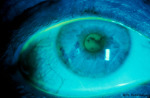
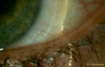
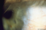
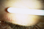
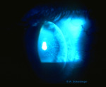
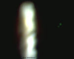
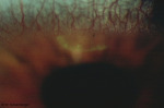
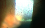
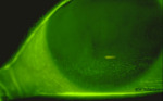
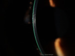
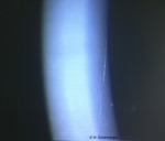
Tear film meniscus
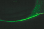
Tear meniscus
Tear meniscus
Tear meniscus
Tear meniscus
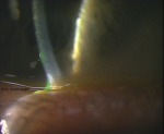
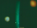
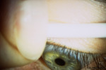
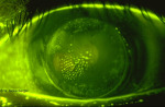
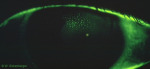
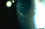
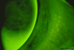
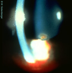
BUT (Break Up Time)
BUT
BUT
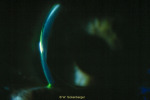
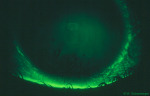
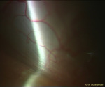
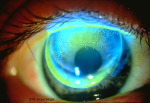
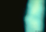
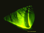
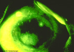
Interference
Interference
Interference
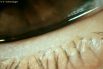
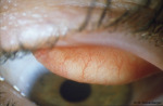
LIPCOF (Lid-Parallel Conjunctival Folds)
LIPCOF
LIPCOF
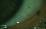
LIPCOF
LIPCOF
Dry eye and contact lenses
Dry eye and contact lenses
Dry eye and contact lenses
Dry eye and contact lenses

Dry eye and contact lenses
Dry eye and contact lenses
Chapter II.1: Tear film -b
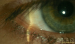
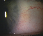
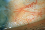
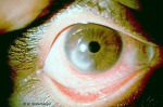
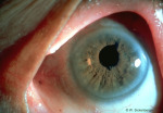
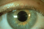
Bulbar conjunctiva
Injection
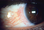
Mixed injection (conjunctival and ciliary)
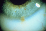
Nevus
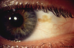
Pigments
Pigments
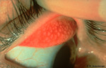
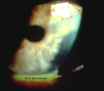
Pinguecula
Nevus
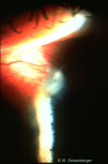
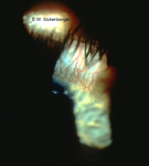
Pterygium
Pterygium
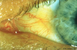
Pterygium
Pterygium
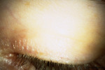
Tarsal conjunctiva
Evert eyelid (Eyelid eversion)
Ectropioning
Ectropioning
Ectropioning
Ectropioning
Ectropioning
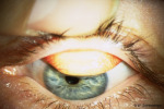
Papillae
GPC
Chapter II.5: Cornea
Epithelium
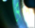
Erosions

Impressions from foreign objects
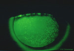
Air bubble dimple veiling
Microcysts / Vacuoles
Mucin balls
SEALS (Superior epithelial arcuate lesions)
Lesions
Cornea
Infiltrates
Optical section
Stroma
Corneal ulcer
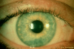
Chapter II.6: Anterior segment findings
images.additional-material.group_2_d41d8
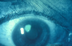
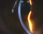
Central depth of the anterior chamber

Anterior chamber
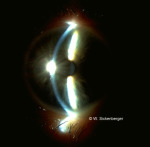
Anterior chamber
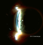
Anterior chamber
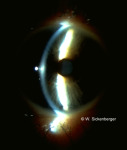
Anterior chamber
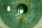
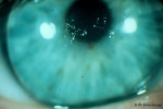
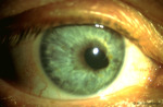
Chapter II.7: Iris
images.additional-material.group_2_d41d8
Central depth of the anterior chamber
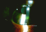
Persisting pupillary membrane (iris strands)
Posterior synechia following iridocyclitis