Case reports
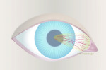
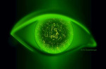
Chapter II.1: Tears Film
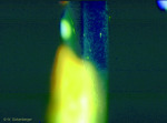
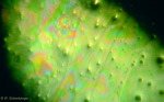
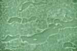
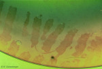
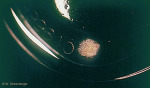
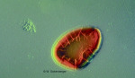
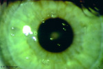
Soft contact lens with dehydrated area
Dryness and contact lenses
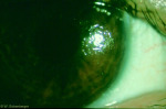
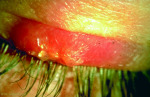
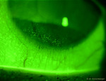
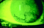
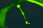
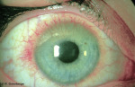
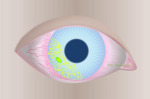
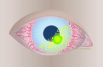
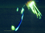
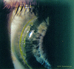
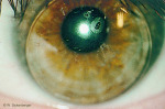
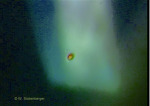
Dry eye disease with a punctal plug (occlusion of the tear duct)
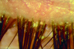
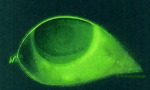
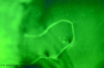
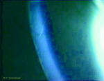
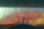
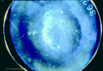
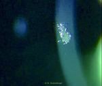
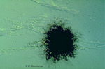
Punctum Plug with high magnification
Chapter II.3: Eyelids
Inflammation of the eyelid (blepharitis) Grade 1
Inflammation of the eyelid (blepharitis) Grade 4
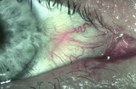
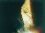
Allergic inflammation of the cutaneous eyelid
Allergic inflammation of the cutaneous eyelid
Incomplete eyelid closure (schematic)
Incomplete eyelid closure
Chapter II.4: Conjunctiva
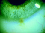
CLIPC - Grade 4
CLIPC - Grade 4
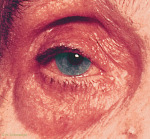
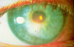
Right Eye: Grade 0
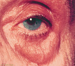
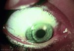
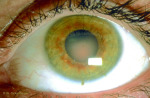
Left Eye: Grade 4
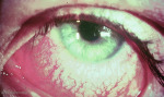
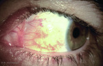
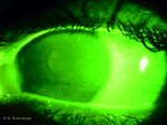
Local conjunctival injection Grade 3
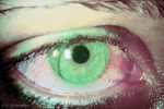
Irritation-free zone conjunctiva superior (same eye)
Conjunctival punctate keratitis
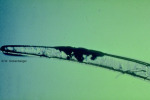
Edge defect in a hydrogel contact lens
Drawing of pinguecula
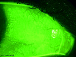
Prominent pinguecula
Drawing of pterygium
Pterygium with injection
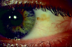
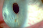
Nevus at 3 o’clock
Nevus in the limbal region at 6 o’ clock
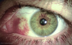
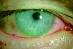
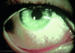
Severe local redness observable in primary eye position
Severe local redness observable when patient looks sideways
Stain from a too tightly fitting soft contact lens
Heavy stain with conjunctival epithelial flap (CEF) from wearing a soft contact lens
Chapter II.5: Cornea
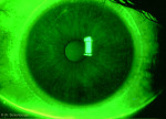
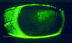
3/9 o’clock punctate keratitis (schematic)
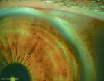
Grade 3 central and peripheral punctate keratitis, confluent at 4 o’clock
Localized punctate keratitis with desiccation (schematic)
Localized punctate keratitis, Grade 2
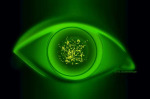
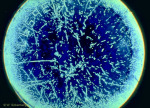
Superficial, diffuse punctate keratitis (schematic)
Diffuse punctate keratitis, Grade 2
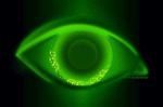
Central punctate keratitis (schematic)
Central punctate keratitis, Grade 3, with impression left by the edge of the contact lens
Deep, diffuse punctate keratitis (schematic)
Grade 4 punctate keratitis due to a toxic reaction
SICS - diffuse distribution
SICS often occurs in connection with punctate keratitis
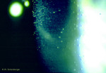
Air bubble dimple veiling (schematic)
Air bubble dimple veiling, without lens
Air bubble dimple veiling beneath rigid contact lenses with fluorescein
Air bubble dimple veiling beneath rigid contact lenses
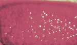
Mucin balls under silicon hydrogel lenses with white light
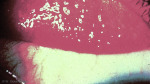
Impressions from mucin balls without contact lenses, after staining with fluorescein
Impression from a foreign object (schematic)
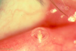
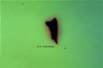
“Scoring” from a foreign object
Imprint of a hair
Deep foreign object impression
SEAL in white light
SEAL after being stained with fluorescein
Erosion (schematic)
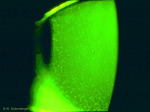
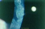
Extensive corneal erosion
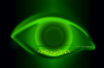
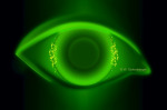
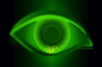
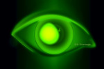
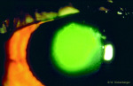
“Halo vision” in cases of corneal oedema
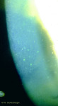
Microcysts and vacuoles, Grade 3
Striae and folds, Grade 3
Individual striae
Pronounced CCC with PMMA contact lenses
CCC after removing contact lenses
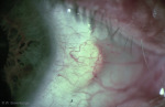
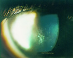
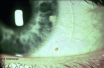
Beginnings of vascularization around 12 o’clock
Massive corneal damage with grade 4 neovascularization
AIK (schematic)
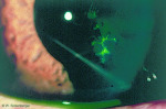
Peripheral infiltrate at 6 o’clock
IK (schematic)
Infiltrate at 5 o’clock with typical limbal injection
MK (schematic)
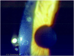
Central corneal ulcer
Active herpes infection with tree-shaped epithelial lesions
Subepithelial scarring zone following secondary infection
Central nummuli following KCE
Central nummuli at high magnification
Central corneal scar of traumatic origin in side view
Angular corneal tear at 5 o'clock with wrinkling
Corneal scars with Descemet’s folds in keratoconus
Corneal scar in grade 4 keratoconus in lateral view
Corneal transplant with lateral observation
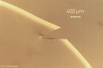
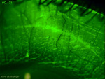
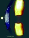
Scars from T-incisions 12 months post surgery (40-fold)
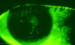
Precipitates in an optical slice
Precipitates in an optical section
Chapter III.3: Deposits
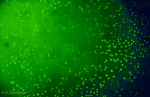
Lipid drops in the tear film (200-fold magnification with endothelial microscope attachment)
Lipid drops on the frontal surface of the lens
Extensive protein deposit, 6-fold magnification, dark field
Netlike protein deposits, 6-fold magnification, dark field
Mucous substances (mucins) on the surface of a hydrogel lens (SEM image)
Deposit at 9 o’clock in the lenticular area of a rigid contact lens (+6.0dpt)
Chapter III.4: Deposits
Hydrophobic areas on rigid contact lenses due to eyeliner residue
Cosmetic residue on a soft contact lens
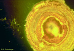
Peripheral metal deposit with discoloration from nicotine, 50-fold magnification, bright field
Central metal deposit with discolouration
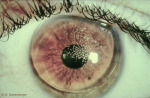
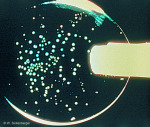
Aspergillus niger, 200-fold magnification, bright field
Aspergillus niger (cross-section of penetration through the contact lens)
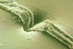
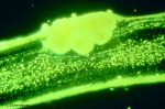
Aspergillus niger, penetration in lens matrix (SEM image)
Candida albicans at 12x magnification
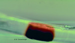
Rust spot on a contact lens
Soft lens, metallic foreign object with rust ring
Rust spot, 200-fold, interference contrast
Cross-section of a contact lens through a rust spot (200-fold magnification)
Metal deposit in the engraving (6-fold magnification)
Metal deposit, 50-fold magnification, bright field
Chapter III.5: Mixed deposits – Jelly bumps
Jelly bumps on a conventional hydrophilic contact lens
Jelly bumps at 6-fold magnification
Top view of a jelly bump
Sectional image of a jelly bump in a cross-section of a contact lens (200-fold)