Schematic Images
Chapter II.1: Tear Film
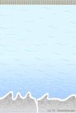
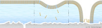
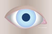
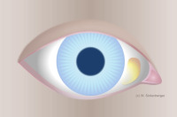
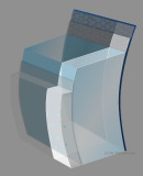
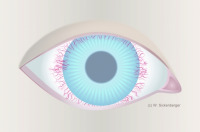
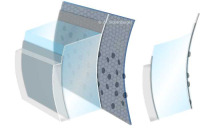
Classical model of the precorneal tear film
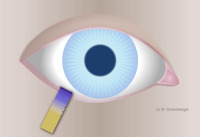
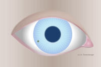
Schirmer test in the temporal third of the lower eyelid
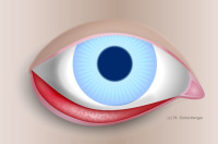
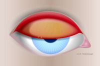
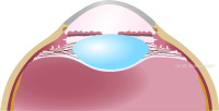
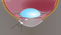
Tear meniscus
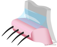
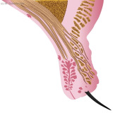
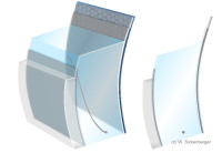
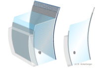
Tear meniscus cross-section
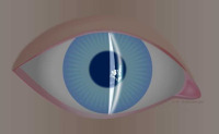
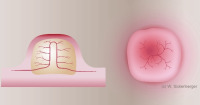
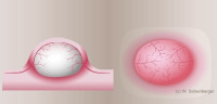
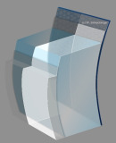
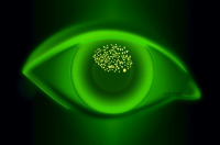
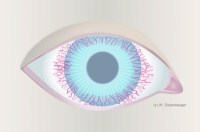
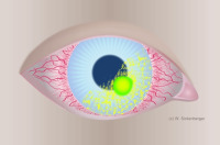
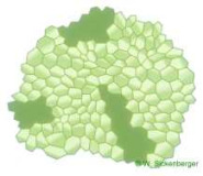
Schematic representation of a ruptured tear film
Dry patches
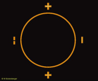
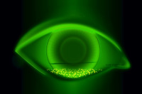
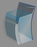
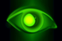
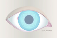
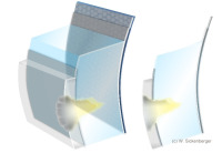
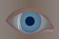
Regular target image of the Sutcliff ophthalmometer in the case of an intact tear film
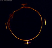
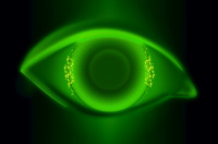
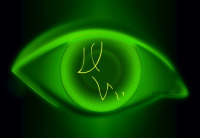
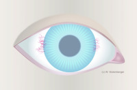
Distorted (anamorphic) Sutcliff ophthalmometer target image in the case of a ruptured tear film
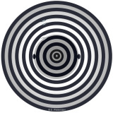
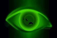
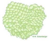
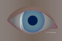
Ideal image of the keratograph targets
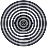
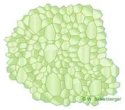
Distorted (anamorphic) image of the keratograph targets

Strongly distorted (anamorphic) image of the keratography targets

Strongly distorted (anamorphic) image of the keratography targets
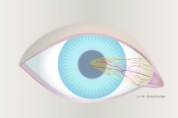
Tear film dynamics
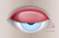
LIPCOF Grade 0
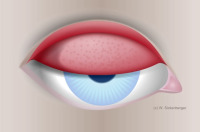
LIPCOF Grade 1
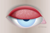
LIPCOF Grade 2
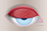
LIPCOF Grade 3
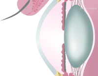
Chapter II.2: Anterior segment findings
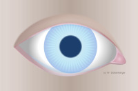
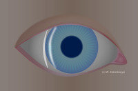
Anterior eye segment
Chapter II.3: Eyelids
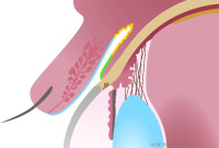
Cross-section of the upper eyelid
Incomplete closure of the eyelid
Chapter II.4: Conjunctiva
Anatomy of the conjunctiva
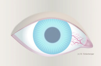
Overview of injection symptoms

Bulbar redness - Grade 0
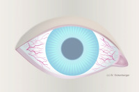
Bulbar redness - Grade 1, Isolated injection
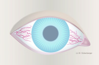
Bulbar redness - Gradе 2
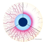
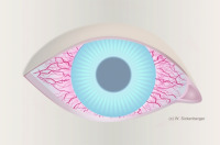
Bulbar redness - Grade 3 bulbar conjunctival injection: 3/9 o’clock redness due to dryness, often associated with 3/9 o’clock superficial punctate keratitis
Bulbar redness - Gradе 4

Everted upper eyelid with papillae
Papillae with central blood vessel
Everted lower eyelid with follicles
Follicle with superficial blood vessels
Grade 0 tarsal conjunctival injection - junctional papillae may be present
Grade 1 tarsal conjunctival injection – slight redness
Grade 2 tarsal conjunctival injection – individual micropapillae (< 0.3 mm)
Grade 3 tarsal conjunctival injection – many macropapillae (0.3 – 1.0 mm)
Grade 4 tarsal conjunctival injection – CLIPC, GPC (papillae > 1.0 mm)
Pinguecula
Pterygium
Chapter II.5: Cornea
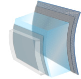
Illustration of the five layers of the cornea
From parallelepiped to optical section

From parallelepiped to optical section
From parallelepiped to optical section

Sclerotic scatter
Optical section for examining the endothelium
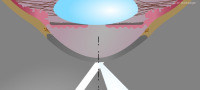
Diffuse illumination
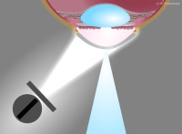
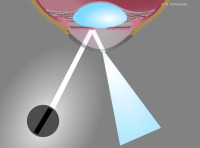
Direct focal illumination: optical plane
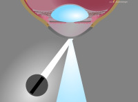
Direct focal illumination: optical cut
Reflective illumination
Conical bundle
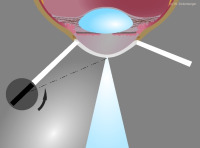
Indirect focal illumination
Retrograde illumination
Sclerotic illumination
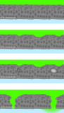
Illustration of various epithelial abnormalities
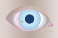
3/9 o’clock punctate keratitis
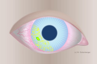
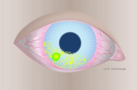
Localized punctate keratitis with desiccation
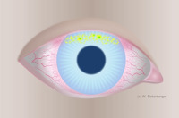
Superficial, diffuse punctate keratitis
Central punctate keratitis
Deep, diffuse punctate keratitis
Air bubble dimple veiling
Impression from a foreign object
Erosion
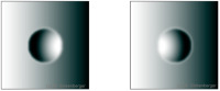
Light-and-shadow phenomenon of microcysts and vacuoles
Location of microcysts and vacuoles

Stromal striae and Descemet’s folds
Location of the striae
Grade 1 vascularization
Grade 2 vascularization
Grade 3 vascularization
Grade 4 vascularization
Location of infiltrates
AI - Asymptomatic infiltrates
AIK - Asymptomatic infiltrative keratitis
IK - Infiltrative Keratitis
CLARE - Contact lens-induced acute red eye
CLPU - Contact lens-induced peripheral ulcer
Corneal ulcer sectional views
MK - Microbial keratitis
Hexagonal arrangement of endothelial cells
Polymegathism
Endothelial blebs
Cornea guttata
Location of precipitates
Chapter II.6: Anterior segment findings
Cross-section through the anterior chamber
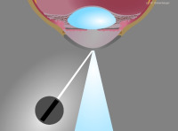
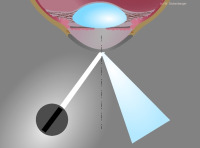
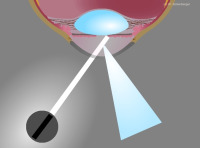
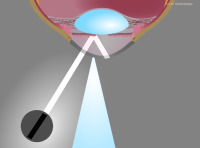
Appearance of the slit image in Van Herick’s method
Measurement setup in Van Herick’s method
Van Herick’s Grade 4 - SC : ACA ratio = 1 : 1
Van Herick’s Grade 3 - SC : ACA ratio = 1 : ½
Van Herick’s Grade 2 - SC:ACA ratio = 1 : ¼
Van Herick’s Grade 1 - SC : ACA ratio = 1 : <¼
Van Herick’s Grade 0 - closed chamber angle
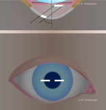
Illustration of Smith’s method – setup
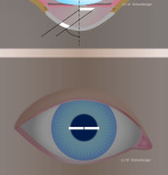
Illustration of Smith’s method - slit images in alignment
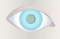
Chapter II.8: The Crystalline lens

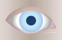
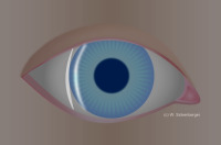
Frontal view
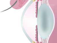
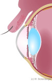
Cross-sectional side view
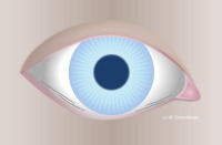
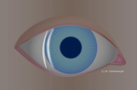
Frontal view
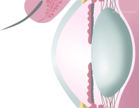
Cross-sectional side view
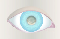
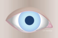
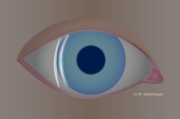
Frontal view
Cross-sectional side view
Chapter IV.4: Appendix (Contact lens terms)
Contact lens terms